
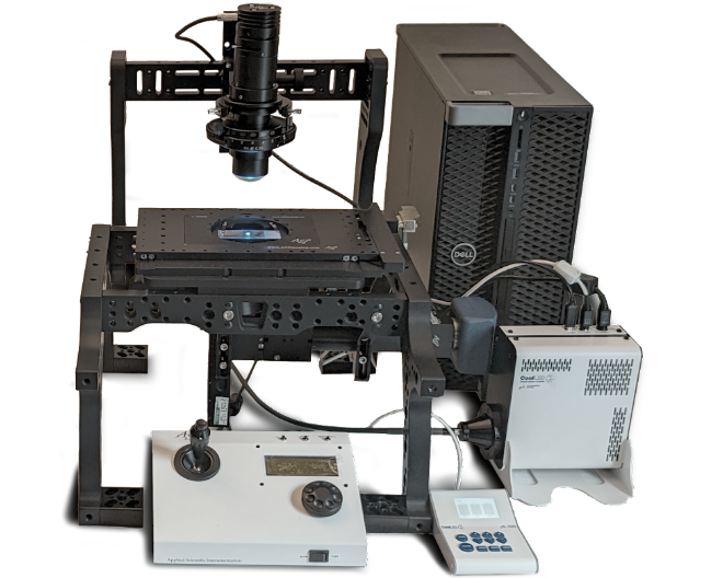
OSTEOIMAGER Side Scanner
The BIOQUANT OSTEOIMAGER slide scanner is an inverted automated scanning microscope. It scans both histology sections and cell cultures. A motorized nosepiece allows it to scan with 4X, 10X, 20X, or 40X objectives. It scans in brightfield, multi-channel fluorescence, polarized light, or darkfield.
Supports BIOQUANT SCAN
BIOQUANT SCAN is the control software for the OSTEOIMAGER. BIOQUANT SCAN is an extension to the BIOQUANT OSTEO or BIOQUANT Life Science analysis software. BIOQUANT SCAN is used to specify objectives, control illumination sources, adjust focus, define scan areas, and export images.
Unified Transmitted / Reflected Light Path
The OSTEOIMAGER slide scanners is designed around a uniform light path that eliminates the need to adjust the microscope when switching between brightfield and fluorescence imaging. Simply turn off one light and turn on the other.
Multi-modal Condenser Wheel
To switch from brightfield to polarized light to darkfield simply rotate the condenser wheel to insert or remove the appropriate filter.
Technical Specifications
Optical Specifications
Olympus UIS2 Fluorite Objectives 4X 0.13NA, 10X 0.25NA, 20X 0.5NA, 40X 0.75NA
Olympus IX2-LWUCD Condenser 25mm Working Distance, 0.55NA
Illumination Specifications
Transmitted Light 10,000 Hour White LED
Epi-fluorescent Light 10,000 Hour Broad Spectrum LED Illuminator
Fluorescent Filter Cube Triple Band ET DAPI/Calcein/Alizarin Red (Changeable)
Polarized Light Linear Polarization, All Objectives
Darkfield Light 4X and 10X Objectives
Sample Handling Specifications
XY Motorized Stage Applied Scientific Instrumentation Model MS2000-FT
Z Motorized Focus Applied Scientific Instrumentation Model LS50
Multi-slide Holder Fixed Vertical Orientation, 4 Slides, 25mm x 75mm
Single-slide Holder 360° Rotation, 1 Slide, 25 x 75mm or 50 x 75mm
Well Plate Holder 1 plate, Standard 84mm x 127mm
Imaging Specifications
Imaging Camera Jenoptik Prokyon - 2.3 / 20 Megapixel, Color, 60fps
Focus Camera Watec 902H3 - 0.3 Megapixel, Monochrome, 30fps
Maximum Scan File Size 4GB Uncompressed
Scan File Formats Calibrated BIF, Uncalibrated TIF
Scan Area 4X over 2500 mm2 (1.4 microns per pixel)
Scan Area 10X over 400 mm2 (0.56 microns per pixel)
Scan Area 20X over 100 mm2 (0.28 microns per pixel)
Scan Area 40X over 25 mm2 (0.14 microns per pixel)
Maximum / Auto Focus Scan Rate 1.5s per field / 5s per field
Imaging Overview
Brightfield Imaging
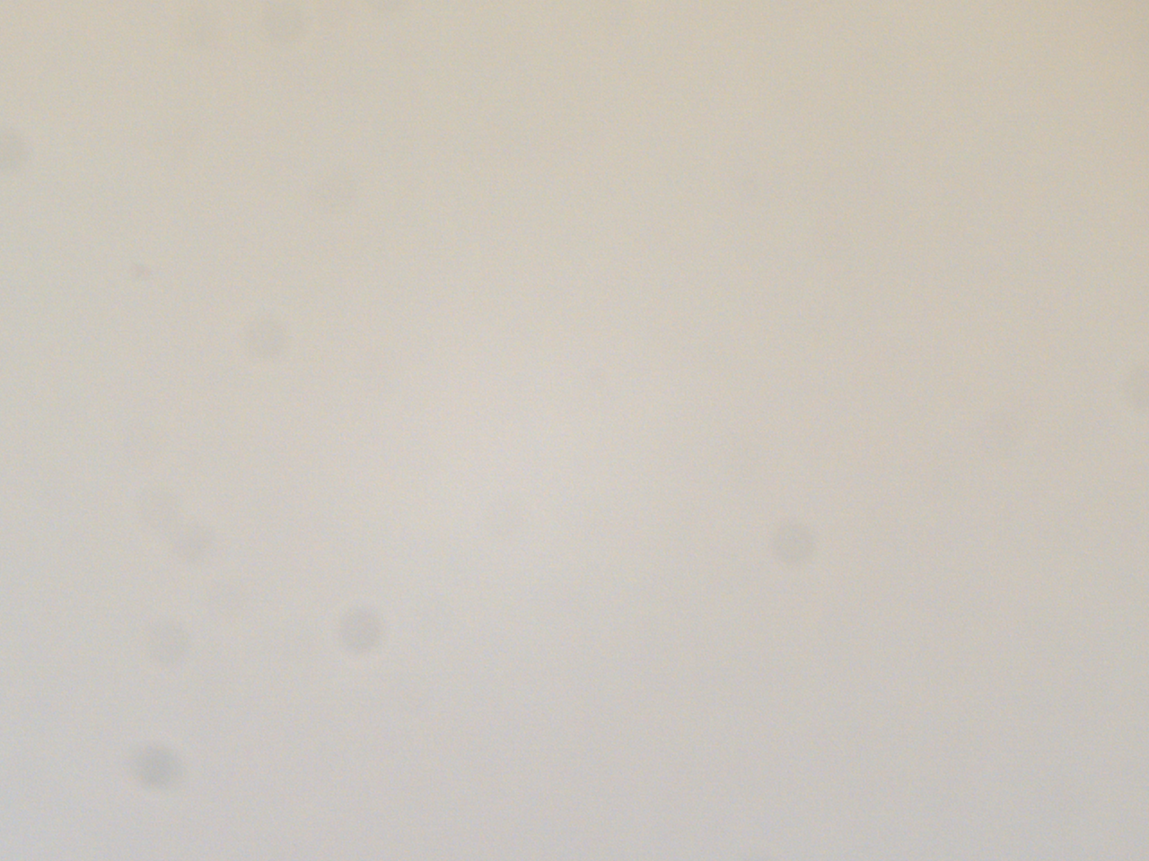
Example of Background Correction
Live Correction dynamically corrects the live image for the uneven lighting.

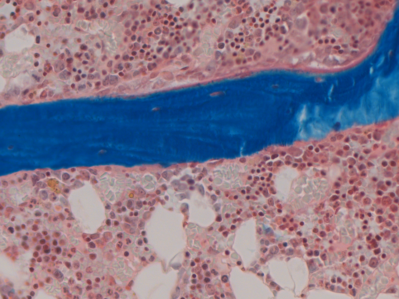
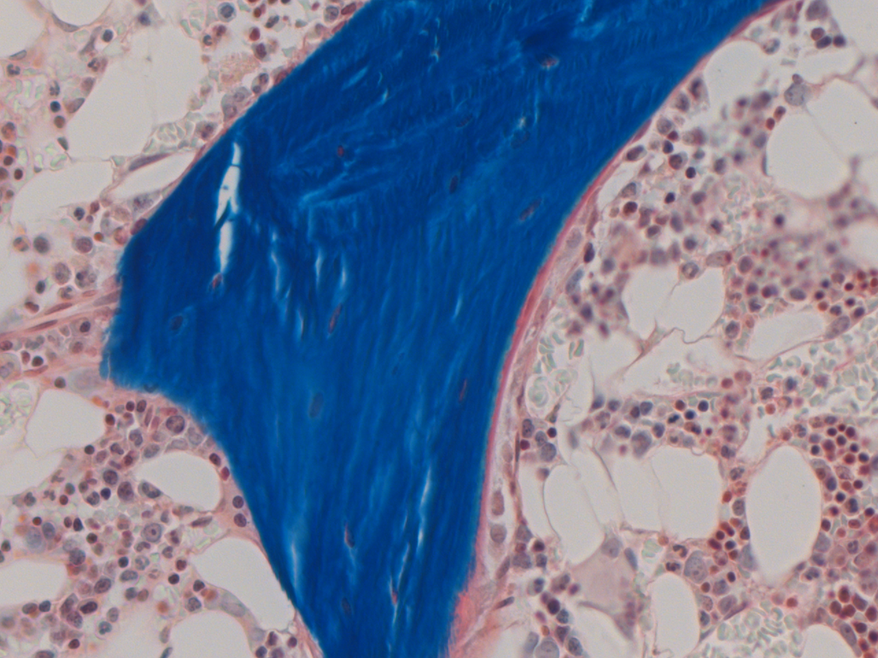
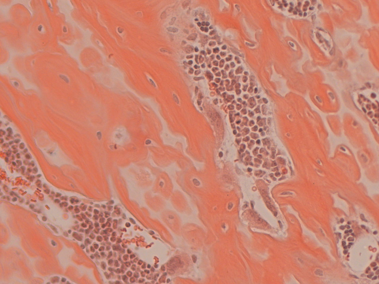
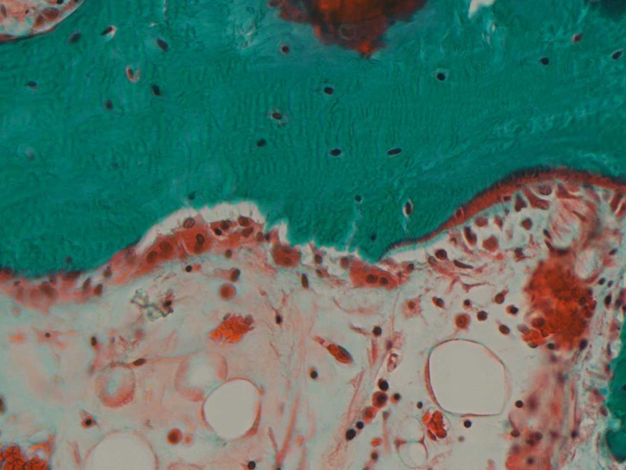
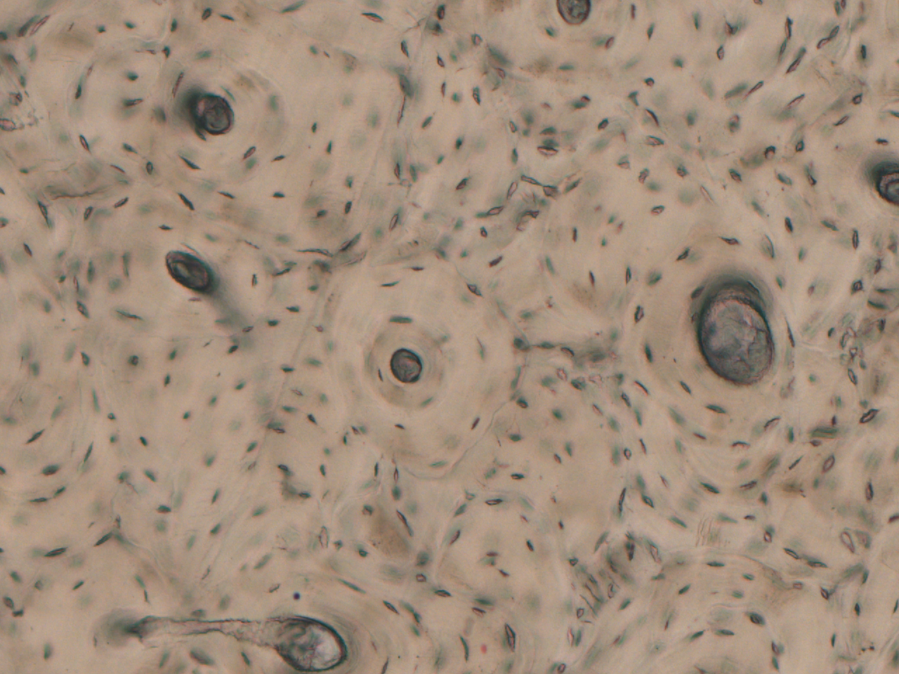
Examples of non-demineralized histology
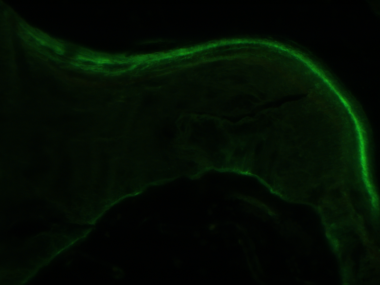
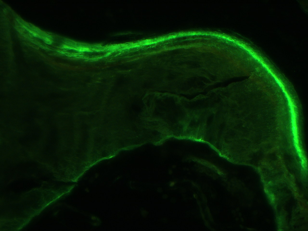
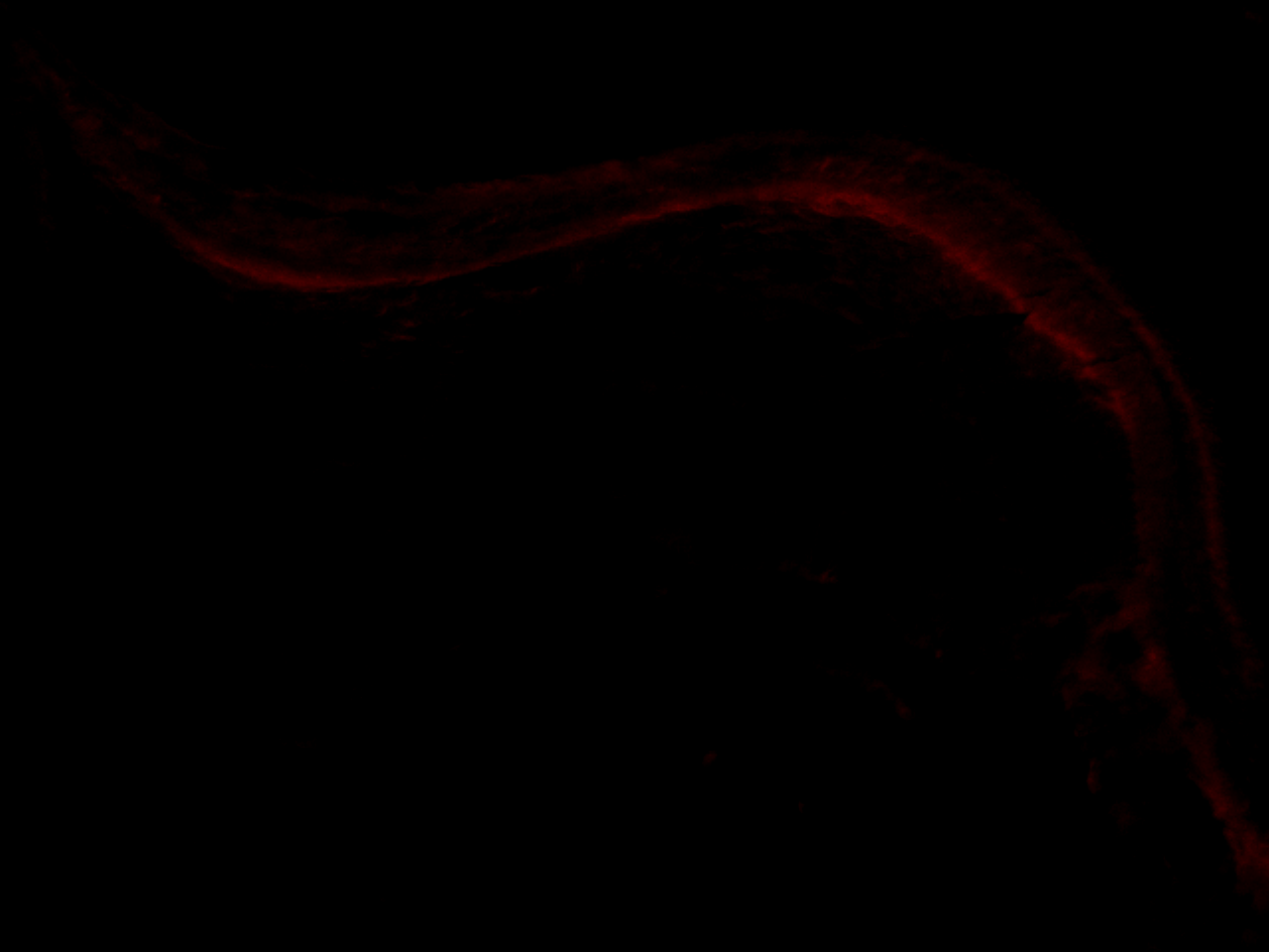
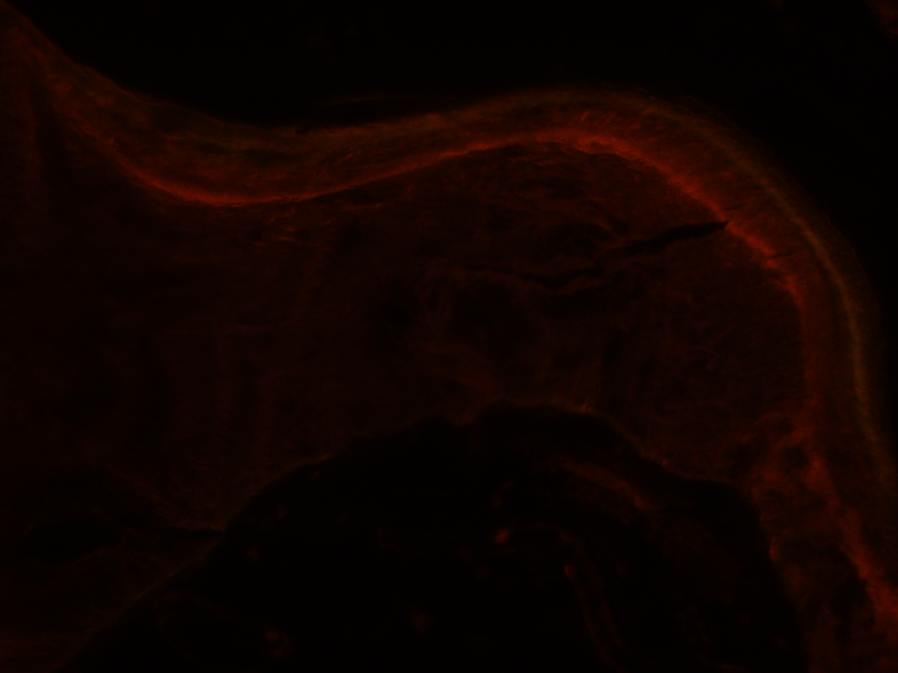
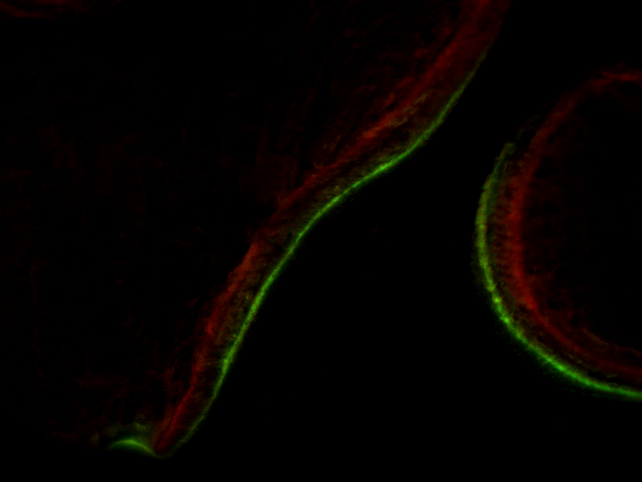
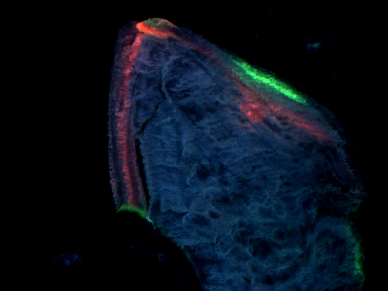
Live Multicolor Fluorescence Mixing
Examples of live multicolor fluorescence mixing with reduced excitation and increased excitation. 20x objective. Calcein and alizarin red labels.
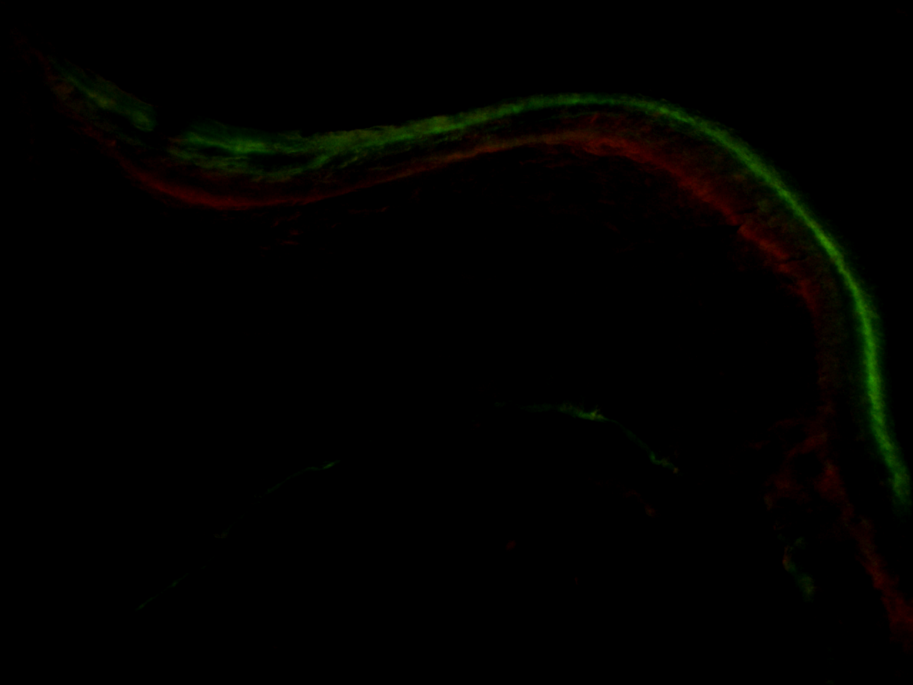
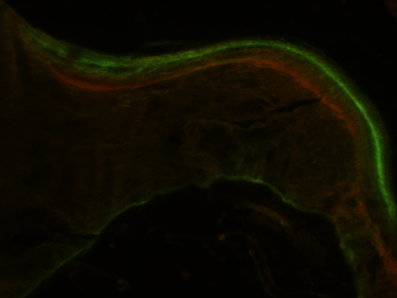
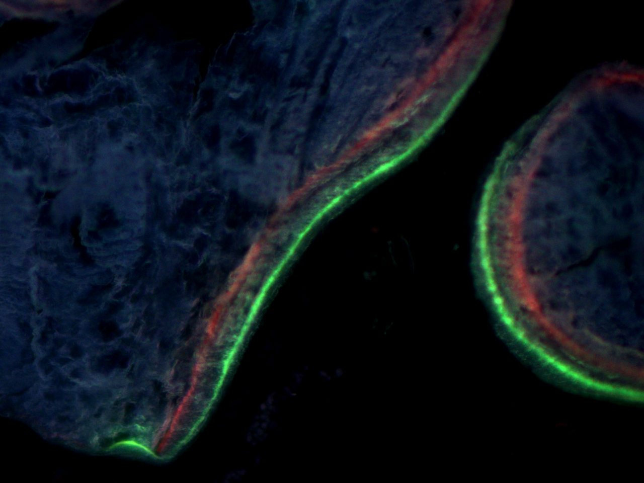
Live Muliticolor Fluorescence Examples
20X objective. 0.4 microns per pixel. Trabecular bone. Calcein and alizarin red labels. Blue autofluorescence in mineralized bone. Hardware brightness adjustment and simultaneous viewing. Live black background correction is also applied in hardware. No software post processing required.

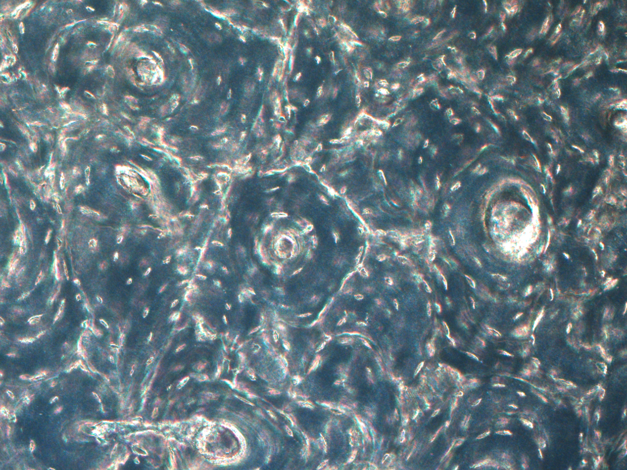
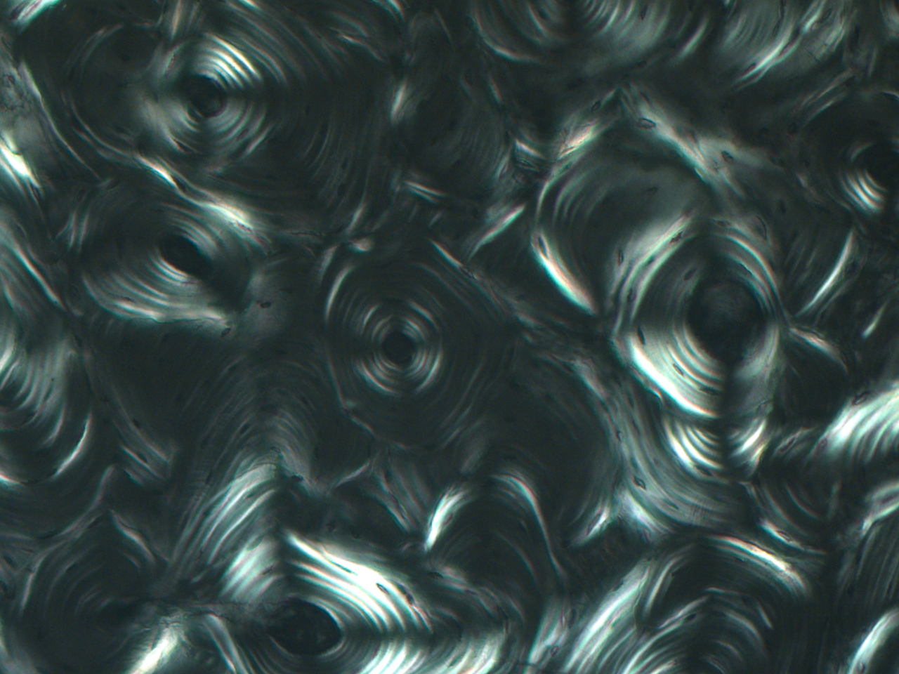
Polarization and Darkfield
Examples of the same field in brightfield, linear polarized illumination, and darkfield illumination.
Sample Scans
Check out sample scans in the Scan Gallery on the BIOQUANT SCAN Add-on page.
Download Data Sheet with Schematic Diagrams
The following data sheet has more information about the OSTEOIMAGER scanning microscope, including schematic diagrams.